Rockford Gastroenterology Associates proudly serves the communities of Rockford, Belvidere, Roscoe, Beloit, and Rockton, Illinois, with expert gastrointestinal care. Through strong collaborations with trusted healthcare partners, Rockford Gastroenterology Associates is dedicated to delivering comprehensive and patient-focused services. Rockford Gastroenterology...read more
With offices in Rockford, Belvidere, Roscoe, and Beloit, Illinois, Rockford Gastroenterology Associates are dedicated to helping patients achieve optimal digestive health. Maintaining a healthy digestive system is essential for overall well-being, as it supports nutrient absorption, immune function, and...read more
Rockford Gastroenterology Associates is excited to partner with Welby Health to offer a new level of personalized, tech-enabled care for our patients. This collaboration reflects our commitment to improving health outcomes through innovation and individualized support. With this partnership,...read more
Finding the best gastroenterologist in Northern Illinois and Southern Wisconsin involves a systematic approach to ensure that you receive high-quality care tailored to your needs. Here is a step-by-step guide to help you identify the best provider in the...read more
Gastroenterologist Dr. Sumeet Tewani was recently featured in Digest Disease Week News article, ‘When to Consider Endoscopic Removal of Ingested Objects’. Here is an excerpt from the article, ‘Gastroenterologists need to recognize the signs and symptoms that require swift...read more
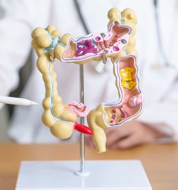
Gastroenterologist Dr. Aaron Shiels was recently featured in 23 WIFR’s news segment, ‘Rockford doctor stresses importance of colorectal cancer screenings’. Here is an excerpt from the article, ‘Colorectal cancer, the second leading cause of cancer death in the U.S.,...read more